Ultrasound


How Does An Ultrasound Help To Diagnose A Breast Lump?
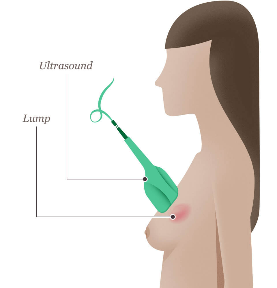
When a suspicious area is detected in your breast through a breast self-exam or on a screening mammogram, your doctor may request an ultrasound of the breast tissue. A breast ultrasound is a scan that uses penetrating sound waves that do not affect or damage the tissue and cannot be heard by humans. The breast tissue deflects these waves causing echoes, which a computer uses to paint a picture of what’s going on inside the breast tissue (no radiation is involved). A mass filled with liquid (which is a benign cyst) shows up differently than a solid mass.
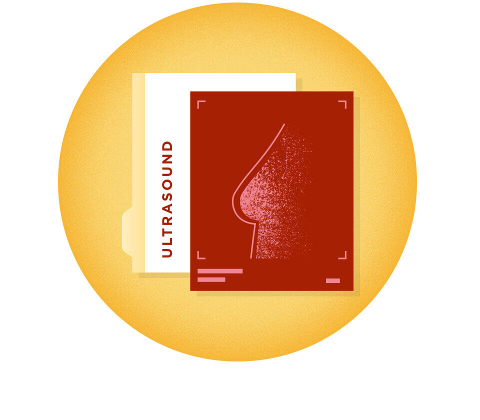
Ultrasound Results: Breast Sonogram
The detailed picture generated by the ultrasound is called a “sonogram.” Ultrasounds are helpful when a lump is large enough to be easily felt, and the images can be used to further evaluate the abnormality.
A breast ultrasound can provide evidence about whether the lump is a solid mass, a cyst filled with fluid, or a combination of the two. While cysts are typically not cancerous, a solid lump may be a cancerous tumor. Healthcare professionals also use this diagnostic method to help measure the exact size and location of the lump and get a closer look at the surrounding tissue. If it is determined that the lump is cancer, the measurement on imaging determines its “clinical” stage.
Abnormal mammogram result? Be informed and ask the right questions.
Si tu mamografía de detección fue anormal, no te alarmes. El recurso gratuito Mamografías Anormales y Qué Hacer Después explica los distintos estudios que podrías necesitar e incluye una lista de preguntas clave para hacerle a tu médico en tu próxima cita.
Prepárate para comprender tus resultados y tomar decisiones informadas sobre los próximos pasos.
Solo disponible en inglés.
¿A dónde te enviamos tu guía gratuita?

Materials on this page courtesy of National Cancer Institute