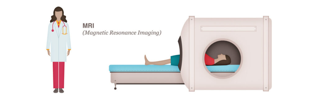
MRI


How does a breast MRI help to diagnose breast cancer?
During diagnostic examinations, it is helpful to get a variety of images and perspectives. If your initial exams are not conclusive, your doctor may recommend a breast MRI (magnetic resonance imaging) to assess the size and specific location within the breast. An MRI can also identify any other abnormal findings within the breast.
During a breast MRI, a magnet connected to a computer transmits magnetic energy and radio waves (not radiation) through the breast tissue. It scans the tissue, making detailed pictures of areas within the breast. These images help the medical team distinguish between normal and diseased tissue.
Dense breast tissue
For women with very dense breast tissue and a family history of breast cancer, it is standard of care to get both a breast MRI and a mammogram annually, rotating these imaging studies every 6 months (mammogram, then 6 months later a breast MRI, then 6 months later a mammogram, etc.).
Radiologists are now required to report the degree of density found within a woman’s breast tissue since highly dense breast tissue is considered a risk factor for developing breast cancer. Having dense breast tissue can make mammogram images harder to read and interpret, which is why additional testing is often required for women with dense breast tissue.
What is dense breast tissue?
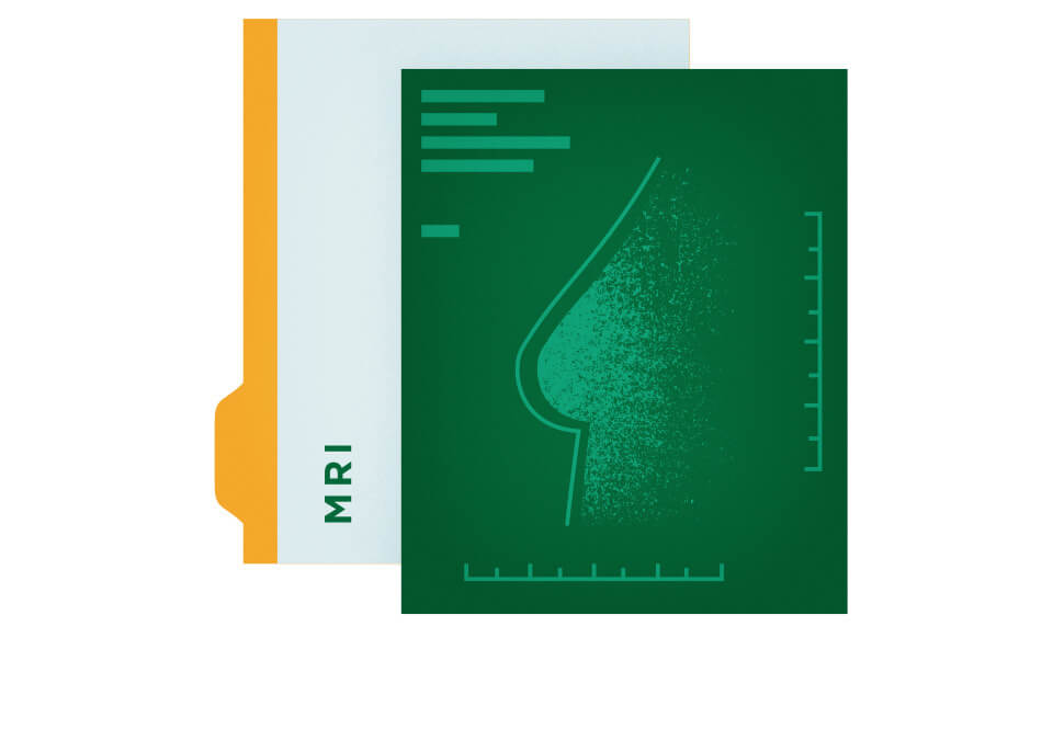
Learn more about dense breast tissue—and what you need to do if you have it—in the free Dense Breast Q&A Guide, written by breast health experts.
Get the Free GuideSources:
Centers for Disease Control and Prevention
National Cancer Institute